
What Is Image Guidance?
Image Guidance is a technique that uses live medical images to help a clinician see body structures during a procedure. The images can show the target area and nearby anatomy. This view helps guide a needle toward the intended joint or tissue during an injection, based on the patient’s anatomy.
Quick Facts About Image Guidance
Category
Procedure guidance technique
Used for
Targeted joint injections
Common confusion
A live guide, not just a scan
Also called
Imaging Guidance, Image-guided procedure
Often discussed with
Joint Injections
Key Takeaways About Image Guidance
- It provides a live view of internal anatomy.
- The clinician guides the needle toward the treatment area.
- It isn’t the same as a diagnostic scan alone.
How Image Guidance Works
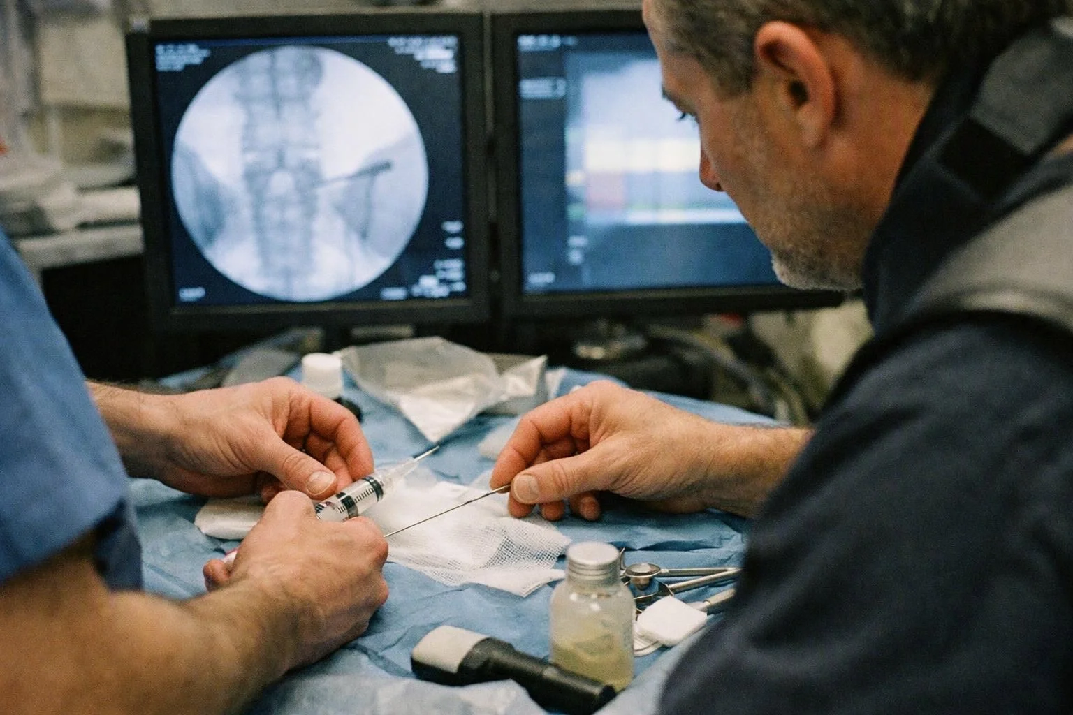
Image Guidance uses a live view of the body to help direct a needle during a joint injection. The clinician reviews the target area on a screen while positioning the needle. This approach connects the planned injection site with the patient’s actual anatomy.
Related glossary terms: Fluoroscopy, Joint Capsule, Bursa.
Ultrasound and fluoroscopy are common imaging methods. Ultrasound creates moving images with sound waves. Fluoroscopy uses X-ray images to show bones and the position of medical instruments. The method depends on the joint, the procedure, and the anatomy being treated.
Image guidance also helps the clinician check needle placement during the procedure. A diagnostic scan taken earlier may show anatomy, but it does not provide the same live view during an injection.
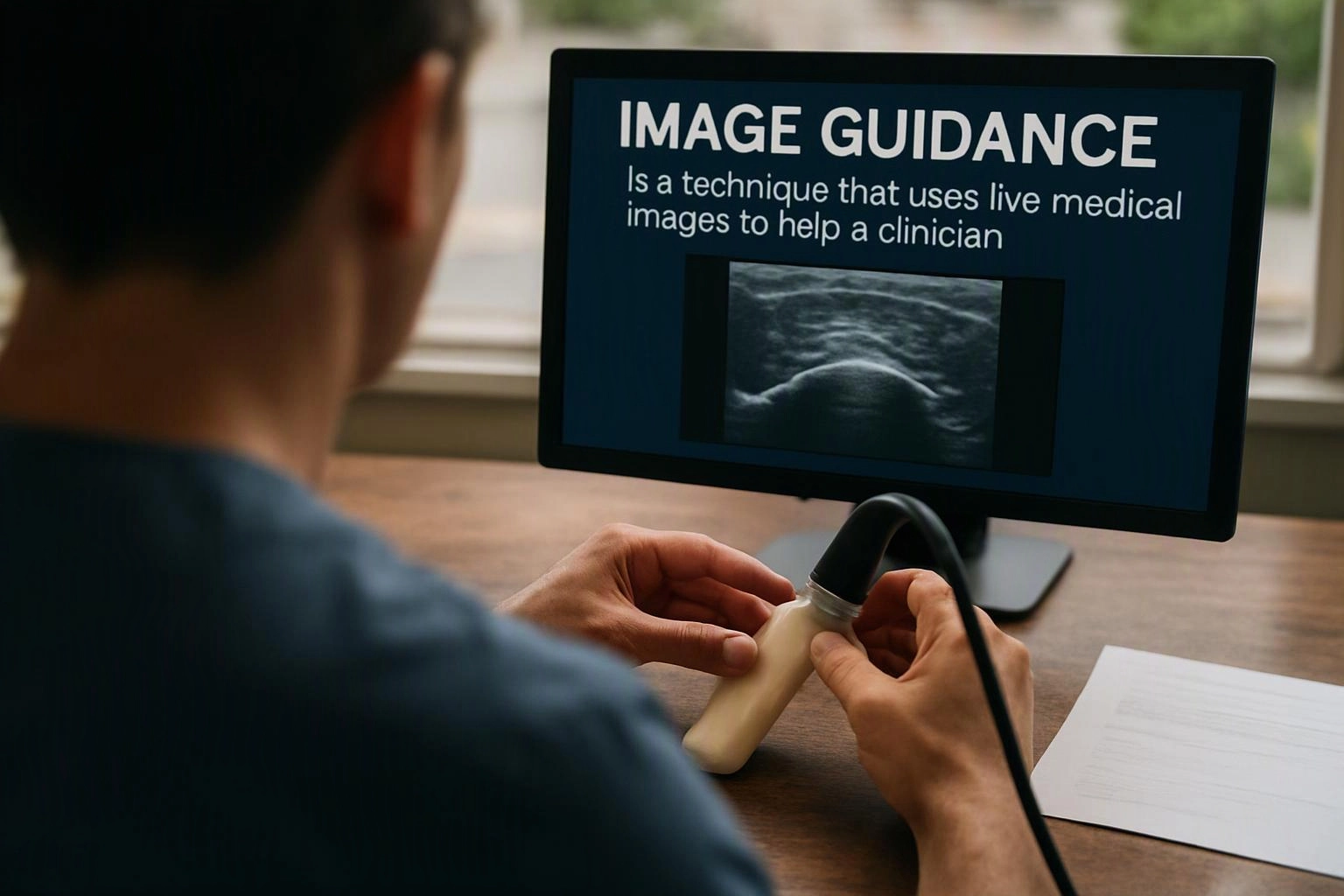
From the Premier Injury Specialists team
When we perform joint injections, we review the target joint and nearby structures before guiding the needle. We watch the image during placement and adjust the path when the patient’s anatomy calls for it. Our team also matches the imaging method to the joint and procedure.
Image Guidance in Practice: A Real-World Example
A patient with knee pain may receive a joint injection with image guidance. The clinician watches the knee on a screen while directing the needle toward the joint space.
Related Terms
Fluoroscopy
Fluoroscopy is a live X-ray imaging method that shows moving body structures on a screen. A…
Joint Capsule
A joint capsule is a tough sleeve of connective tissue that surrounds a movable joint. The…
Bursa
Bursa is a small, fluid-filled sac near a joint that reduces rubbing between bones, tendons, muscles…
Facet Joint
A Facet Joint is a small connection between the bones at the back of the spine…
Premier Injury Specialists
Have Questions About Image Guidance?
Contact Premier Injury Specialists for practical guidance on Image Guidance and related pain management physician work in Stonecrest.